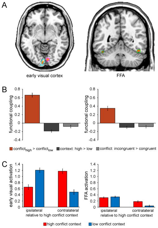
Figure 4.
Context-specific improvements in interference resolution are mediated by top-down control. A, Functional coupling with the right mSPL as a function of context-specific variation in conflict processing (interferencehigh-conflict context > interferencelow-conflict context) as revealed by PPI analysis is shown for voxels in the independently-localized early visual cortex [left panel; left hemisphere (light blue outlines): x -2, y -86, z -12; 290 voxels; right hemisphere (magenta outlines): x 10, y -80, z -8; 75 voxels] and FFA ROIs (right panel; green outlines; left hemisphere: x -44, y -46, z -26; 35 voxels; right hemisphere: x 48, y -56, z -22; 53 voxels), displayed at p < .05 (small volume correction) on axial (z -7) and coronal (y -50) slices of an individual brain in MNI space. B, Greater functional coupling (beta estimates ± SEM) between the mSPL and bilateral early visual cortex (left panel) and FFA (right panel) as a function of context-specific variation in conflict processing (orange) relative to that as a function general context representation (dark gray) and conflict processing (light gray) as revealed by PPI analysis (one-way ANOVAs: both F(1,24) > 29.1, both p < .0001). C, Mean activation (beta estimates ± SEM) in the early visual cortex (left panel) and FFA (right panel) ROIs is plotted as a function of the laterality of the anatomical hemisphere (relative to the high-conflict location/visual hemifield: ipsi- vs. contralateral) and context (high conflict vs. low conflict). Note that activation is not plotted relative to stimulus presentation per se given the counterbalancing of the contextual conflict frequency manipulation across participants.

