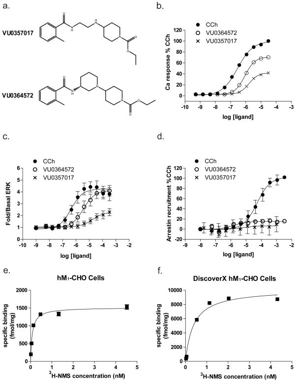
Figure 2. VU0364572 and VU0357017 induce calcium release and ERK phosphorylation but are without effects on β-arrestin recruitment.
(a.) Chemical structures of the two M1 agonists VU0357017 and VU0364572. (b.) Concentration response curves (CRCs) of receptor-induced calcium release for CCh (filled circles), VU0364572 (open circles), and VU0357017 (crosses) in CHO-K1 cells stably expressing human M1 mAChRs. Data are normalized to the CCh maximum response. Data points represent mean ± S.E.M of four independent experiments preformed in duplicate or triplicate. (c.) CRCs of agonist-induced ERK1/2 phosphorylation (pERK1/2) assessed using the SureFire ERK phosphorylation assay in hM1 CHO cells. Data is expressed as fold change over basal ERK levels and is normalized to the maximum response elicited by CCh. Data represent the mean ± S.E.M. of 7–8 independent experiments performed in duplicate or triplicate. (d.) CRCs of agonist-induced β-arrestin recruitment in hM1 CHO cells using PathHunter detection kit. Data points represent mean ± S.E.M. of three independent experiments performed in duplicate or triplicate and are normalized to %CCh max. (e.) Saturation isotherms of [3H] NMS binding to membranes prepared from hM1 CHO cells. Receptor density values (1479 ± 129 fmol/mg protein) were obtained from three independent experiments. (f.) Representative saturation isotherms of [3H] NMS binding to membranes prepared from hM1 CHO cells used in β-arrestin recruitment assays. Receptor density values (11701.600 ± 1411.21 fmol/mg protein) were obtained from five independent experiments.

