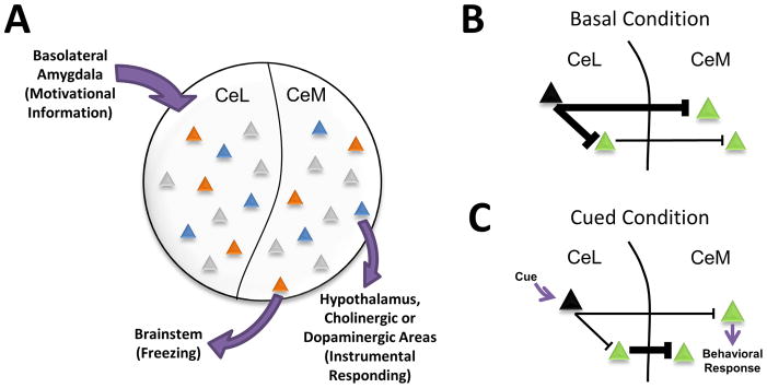
Figure 1. A Model of Behavioral Selection by Neurons in the Central Amygdala.
A. Cues associated with different types of aversive stimuli are represented in distinct patterns of neural activity (indicated by different colored triangles) within the central nucleus of the amygdala (CeA). Salient information about the cues is relayed to the CeA via afferents from the basolateral amygdala. Cues paired with different forms of aversive stimuli, such as shock (orange) or omission of expected reward (blue), recruit non-overlapping populations of neurons in both the lateral (CeL) and medial (CeM) subdivisions of the CeA (Purgert et al., 2012). We hypothesize that these networks of coactive neurons not only represent distinct aversive cues, but also target different downstream structures in order to select appropriate behavioral responses. B, C. A general model by which both subregions of the CeA may be recruited by aversive cues. Under basal conditions (B), cue-inhibited neurons (black) provide tonic inhibition throughout CeL/CeM microcircuits involved in behavioral expression. Upon cue presentation (C), inhibition of these neurons results in the disinhibition of projection cells (green) in both the CeL and CeM, allowing for activity across both subnuclei and the selection of an appropriate behavioral response. (Orange triangles – shock cue activated neurons; Blue triangles – omission cue activated neurons, Black triangles – cue inhibited neurons, Green triangles – projection neurons)

