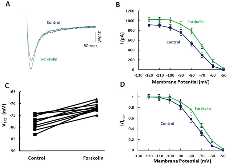
Figure 3. cAMP modulation of Ih tail current.

A. Individual tail currents were measured in control (blue) and after 10 minute bath application of 50μM forskolin (green). B. Population averages demonstrate that forskolin significantly increases the tail current measured at −40mV after stepping from a series of holding potentials (−120mV to −50mV). C. The V1/2 values, generated from Boltzman fits to the normalized activation curves, are measured across individual cells for control (square) and for forskolin (triangle). D. Tail current activation curves obtained in control (blue square) and in the presence of forskolin (green triangle) are shown normalized to maximum current.
