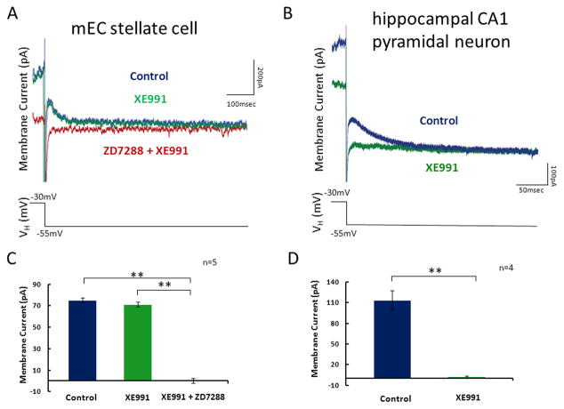
Figure 4. Analysis of IM in stellate neurons and CA1 pyramidal neurons in the hippocampus.
A. Whole cell patch clamp recordings were made in mEC layer II stellate cells. Tail currents measured during voltage steps to −55mV from a holding potential of −30mV in control ACSF (blue), after application of XE991 (green) and finally after application of XE991 with ZD7288 (red). B. Populations averages demonstrate that there is no significant change in the membrane current response between control ACSF and XE991. After subsequent application of ZD7288 the rectifying membrane current is completely abolished. C. Whole cell patch recordings were made in hippocampal CA1 pyramidal neurons. Tail currents were measured in control ACSF (blue) and after application of XE991. Tail currents measured during voltage steps to −55mV from a holding potential of −30mV in control ACSF (blue) and after application of XE991 (green). D. Populations averages demonstrate that application of XE991 removes the membrane current.

