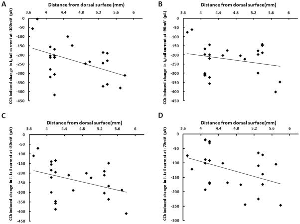
Figure 5. Cholinergic modulation of Ih tail current differs along the dorsal-ventral axis of medial entorhinal cortex.
A. Changes in tail current amplitudes induced by carbachol (CCh) at steps from a holding potential of −100mV to −40 mV, plotted against the dorsal-ventral anatomical location of the stellate cell relative to the dorsal surface of the brain. The linear fit demonstrates significantly larger cholinergic modulation of the Ih tail current in ventral mEC (p=0.024, n=24). B. For changes in tail current amplitudes induced by carbachol at steps from −90mV to −40 mV, the linear fit demonstrates a trend towards larger cholinergic modulation of the Ih tail current in ventral mEC (p=0.268, n=24). C. Changes in tail current amplitudes measured at steps from −80mV to −40mV show a significantly larger cholinergic modulation of the Ih tail current in ventral mEC (p=0.041, n=24). D. Changes in tail current amplitudes measured at steps from −70mV to −40mV show a significantly larger cholinergic modulation of the Ih tail current in ventral mEC (p=0.0467, n=24).

