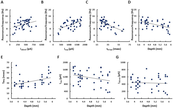
Figure 7. Analysis of the baseline electrophysiological properties of stellate cells.
A. The magnitude of Ih tail current measured after voltage steps from −80mV to −40mV demonstrates a significant positive relationship with membrane potential resonance frequency (p=0.0354, n=39). B. Ih tail current measured at voltage steps from −120mV to −40mV demonstrates a significant positive relationship with membrane potential resonance frequency (p=0.0007, n=39). C. The time constant of Ih activation measured at voltage steps from −80mV to −40mV demonstrates a significant positive relationship with membrane potential resonance frequency (p=0.0001, n=36). D. The membrane resonance frequency decreases significantly from dorsal to ventral mEC (p=0.0117, n=36). E. The time constants of Ih activation increases significantly from dorsal to ventral mEC (p=0.0117, n=36). F. The maximum Ih tail current measure at voltage steps from −120mV to −40mV shows a trend towards larger amplitude in dorsal mEC, but this does not reach the criteria for statistical significance (p=0.081, n=39). G. There is no significant relationship between Ih tail current amplitude and dorsal-ventral location for tail currents measured at −80mV (p=0.7197, n=39).

