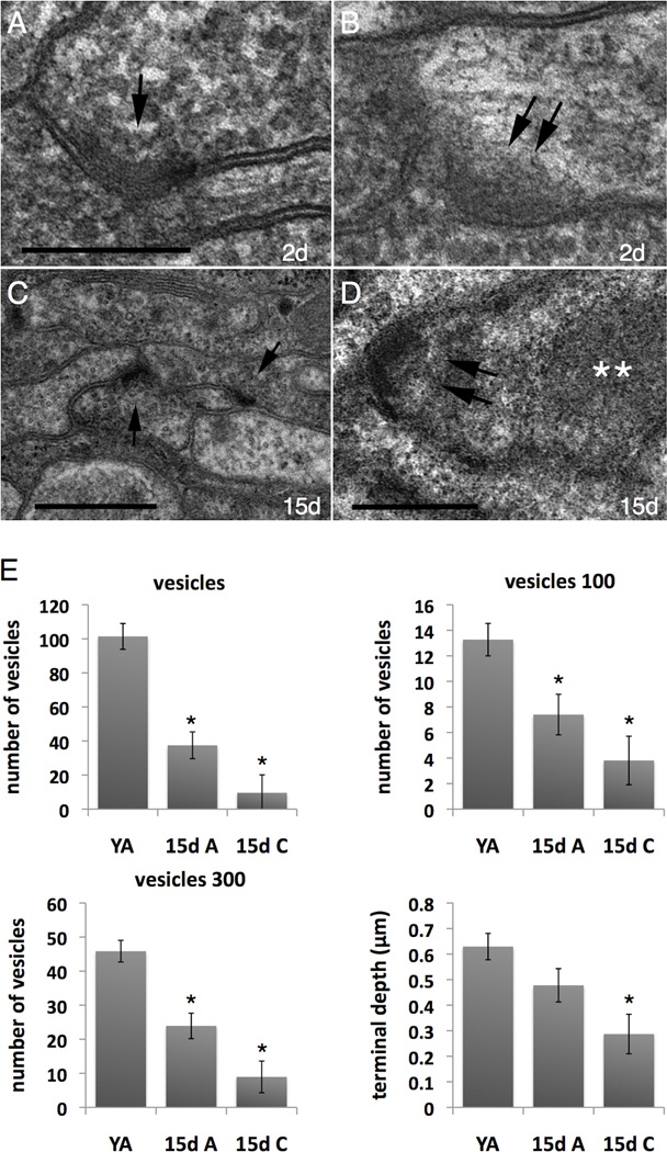
Figure 6. C. elegans synapses deteriorate with age, with extent of synaptic reduction correlated with degree of mobility impairment.
A. Closeup of a normal synapse (arrow) in a young adult animal. A prominent presynaptic bar lies along the plasma membrane, and the process is swollen with synaptic vesicles. Vesicles lying close to the bar are somewhat smaller in diameter than vesicles away from the release zone. There is no anatomical specialization seen on the postsynaptic process. Scale bar is 0.25 microns for panels A & B.
B. A depleted synapse (double arrows) in the same young adult displays a normal presynaptic bar, but a paucity of synaptic vesicles close to the bar or at a distance.
C. In a Class A adult at 15 days of adulthood (20°C), chemical synapses (arrows) remain well organized but have fewer vesicles near the presynaptic bar and the presynaptic process is therefore smaller in diameter. Note that many nearby axons (away from the synapse) remain almost the same diameter as in a young adult. Many axons still contain clusters of synaptic vesicles and small bundles of microtubules. Scale bar is 0.5 microns.
D. Closeup of a depleted synapse (double arrows) in a Class A animal at 15 days of adult life. A fuzzy electron dense inclusion (white asterisks) lies close to the depleted synapse. This may represent pathological deposition of cytoplasmic proteins. Scale bar is 0.25 microns.
E. Quantitation of synaptic features in aging C. elegans. YA, young adult; 15d A, 15 day old class A animal (20°C) that is relatively vigorous for its same-age counterparts and considered to have aged gracefully; 15d C, 15 day old class C animal that is decrepit, barely mobile, and considered to have aged poorly. Data include measurements of 51 synapses from 6 young adults; 52 synapses from 3 Class A animals; 28 synapses from 3 Class C animals. Synapses were from the nerve ring and lateral ganglia. “Vesicles” indicates counts of all vesicles; “vesicles 100” counts all within 100 nm of the synaptic density; “vesicles 300” scores all within 300 nm from the synaptic density. Asterisks indicate p<0.02 as compared to young adult values; repeated measures analysis of variance test (SAS program). For terminal length, YA compared to 15d A, p=0.10. See notes on measuring terminal length in Methods.

