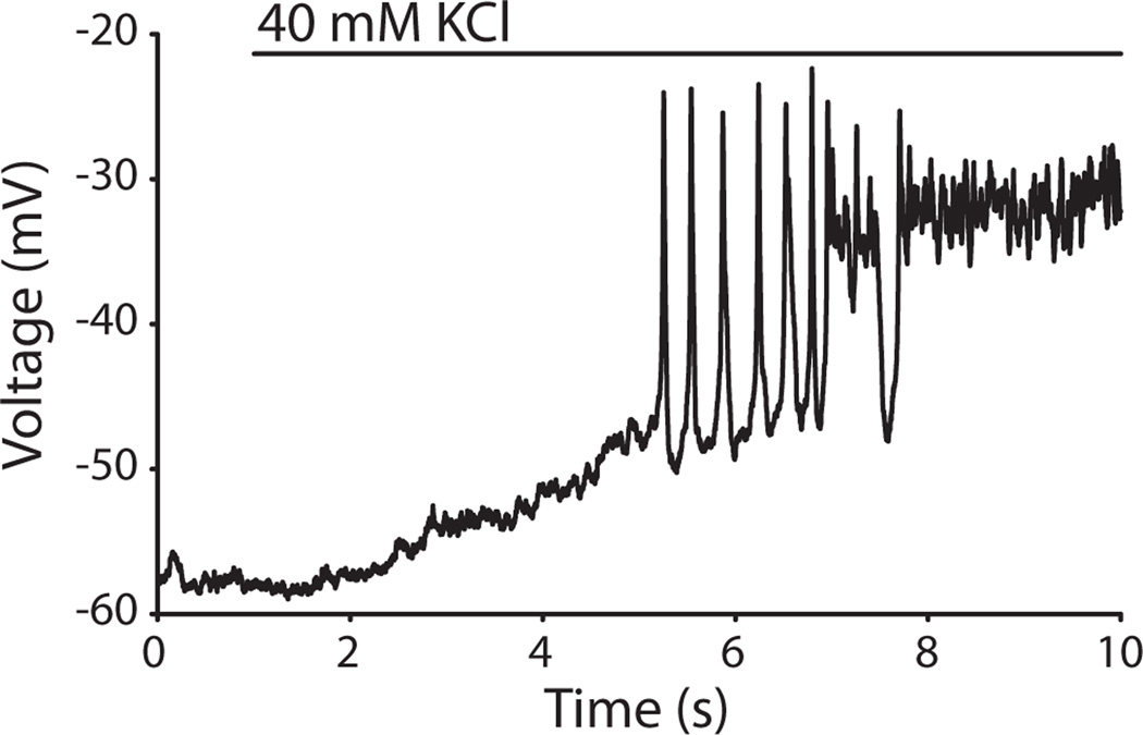
Figure 8.
OHC depolarization by ‘whole-field’ application of high potassium saline. Representative OHC recording shown. A large bore perfusion pipette was used to apply 40 mM potassium extracellular solution to the tissue while performing whole-cell current-clamp recordings from an OHC. The OHC responded with a slow depolarization, followed by an average of 8 spikes (n=4 OHCs). The OHC membrane potential then reached a plateau depolarization for the remainder of the high potassium application.

