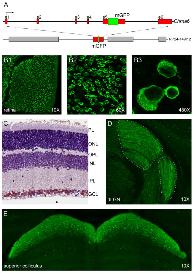
Figure 1.
Production of α6-GFP BAC transgenic mice and α6* nAChR expression in visual system. A. Construction of α6-GFP bacterial artificial chromosome (BAC) transgene. A BAC containing the full Chrna6 gene was modified by recombineering to allow expression of a variantα6 nAChR subunit with GFP fused in-frame within the M3–M4 intracellular loop. B, C. α6* nAChR expression in retina. Whole-mount retinal sections were prepared from α6-GFP transgenic mice and stained with anti-GFP antibodies. GFP(+) cells in the ganglion cell layer (GCL) were imaged with confocal microscopy at 10X (B1), 60X (B2), and 480X (B3). Retinal cross sections were prepared from α6-GFP transgenic mice (C), followed by anti-GFP staining (brown color) and hematoxylin counterstaining to label nuclei. α6 immunoreactivity was confined to the RGC layer. D, E. α6* nAChR expression in visual thalamus and superior colliculus (SC). Brains from α6-GFP transgenic mice were fixed, sectioned at 50 μm (coronal), and stained with anti-GFP antibodies. Dorsal lateral geniculate nucleus (dLGN) (D) and SC (E) stained positive for α6* nAChR expression.

