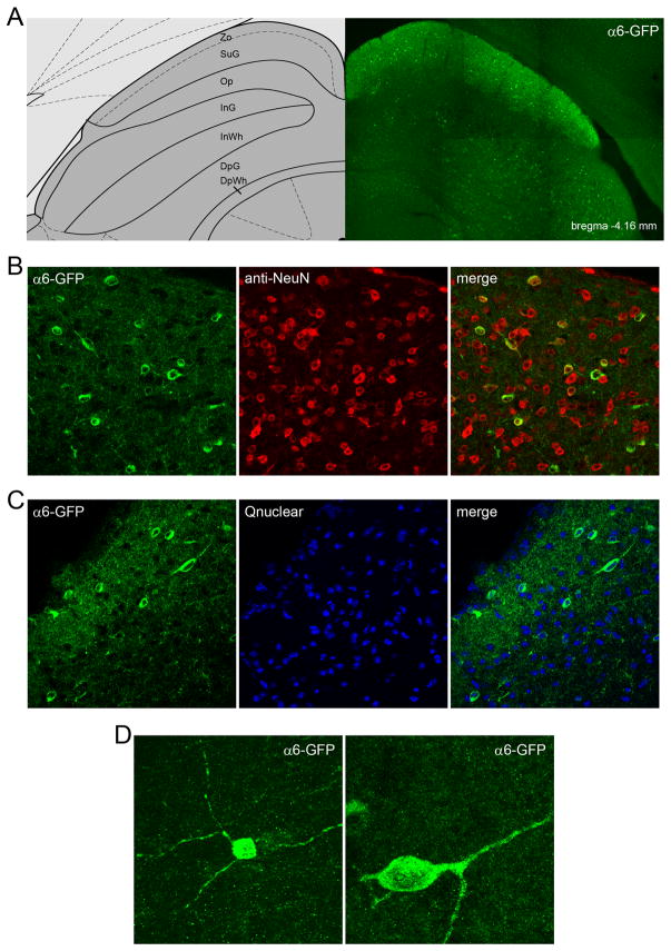
Figure 4.
α6* nAChR expression in superficial superior colliculus neurons. A. α6* nAChR expression in a GFP-stained coronal section (bregma −4.16 mm) from α6-GFP transgenic mice is shown next to a diagram of the SC (Paxinos and Franklin, 2001). α6* nAChR expression is limited to the zonal layer (Zo), the superficial gray layer (SuG), and intermediate gray layer (InG). B, C. α6* nAChRs are expressed in a subset of neurons in sSC. In B, SC coronal sections were double stained with anti-GFP and anti-NeuN antibodies, followed by laser scanning confocal imaging. Individual GFP and NeuN channels and a merge micrograph are shown, at 60X magnification. In C, similar sections were double stained with anti-GFP antibodies and a nuclear stain, followed by confocal imaging as in B. D. Z-stack rendering of α6(+) neurons in sSC. Two α6(+) neurons in sSC were imaged with confocal microscopy and a 3D volume render of the cell body and its processes was created following serial Z-sectioning through the neuron.

