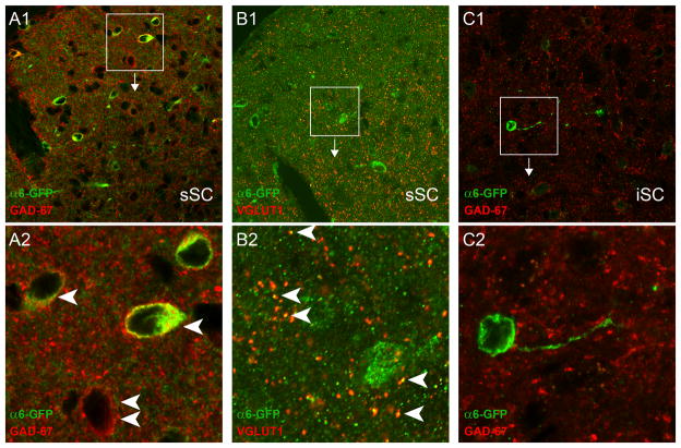
Figure 5.
Transmitter phenotypes of α6(+) neurons in superior colliculus. A. α6* nAChRs are expressed in GABAergic neurons in sSC. Coronal sections from α6-GFP transgenic mice containing sSC were double stained with anti-GFP and anti-GAD67 antibodies, followed by confocal microscopy. A2 represents the boxed area in the A1 image. Single white arrows indicate cells co-expressing α6* nAChRs and GAD67, whereas double white arrows indicate cells expressing only GAD67. B. α6* nAChRs are also expressed in glutamatergic fibers in sSC. Coronal sections from α6-GFP transgenic mice containing sSC were double stained with anti-GFP and anti-VGLUT1 antibodies, followed by confocal microscopy. B2 represents the boxed area in the B1 image. White arrows indicate co-localized VGLUT1 and α6* nAChRs. C. Intermediate superior colliculus (iSC)α6(+) neurons are not GABAergic. Coronal sections from α6-GFP transgenic mice containing iSC were double stained with anti-GFP and anti-GAD67 antibodies, followed by confocal microscopy. C2 represents the boxed area in the C1 image.

