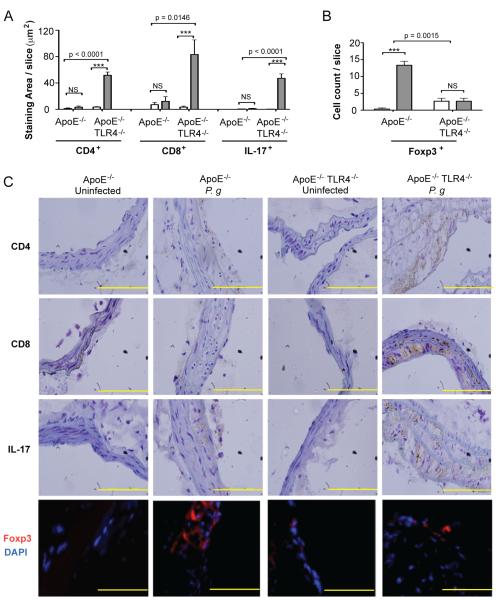
Figure 4. TLR4 deficiency promotes Th17/Treg imbalance in atherosclerotic lesions following infection with P. gingivalis.
Quantitative immunohistochemistry of (A) CD4, CD8, IL-17 positive staining area in the innominate artery of uninfected and P. gingivalis infected ApoE−/− and ApoE−/−TLR4−/− mice using ImageJ software (NIH) or (B) Foxp3 positive cell count. Three sections from n=4 mice/group were analyzed. White bar: Uninfected. Gray bar: P. gingivalis infected. Data are mean ± SD of positive staining area/slice or cell count/slice. Intra-genotype comparisons were calculated by Mann Whitney U test ***p<0.0001. NS= not significant. Inter-genotype comparisons were calculated by Two-way ANOVA (indicated p values). (C) Representative immunohistochemistry of CD4, CD8, IL-17, and Foxp3 positive cells in the innominate artery of uninfected and P. gingivalis infected ApoE−/− and ApoE−/−TLR4−/− mice. Scale=5μm.

