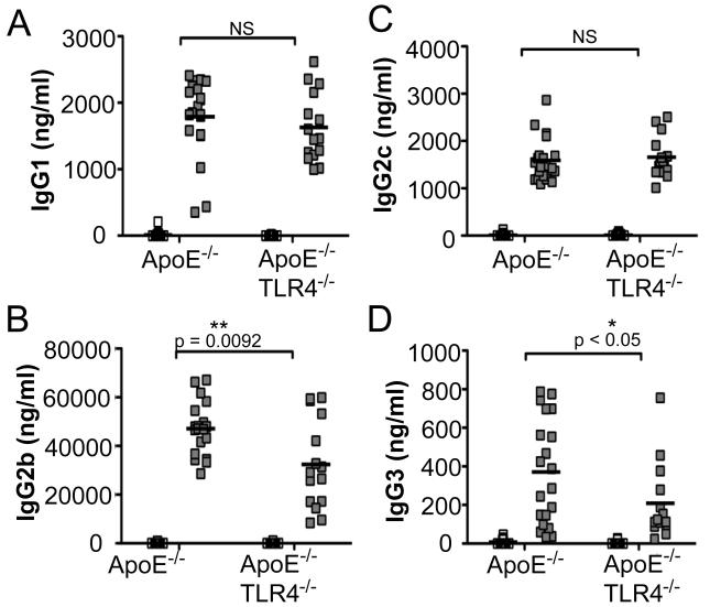
Figure 5. P. gingivalis-specific antibody isotypes IgG1, IgG2b, IgG2c, and IgG3.
P. gingivalis-specific IgG production in uninfected ApoE−/− (n=15), P. gingivalis infected ApoE−/− (n=21), uninfected ApoE−/−TLR4−/− (n=17) and P. gingivalis infected ApoE−/−TLR4−/− (n=14) mice as measured by ELISA. (A) IgG1, (B) IgG2b, (C) IgG2C, (D) IgG3. White squares= uninfected, Gray squares= P. gingivalis infected. Data were analyzed by Student’s t-test. *p<0.05, **p=0.0092. NS=not significant.

