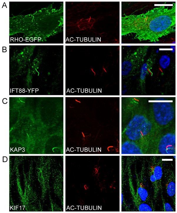
Fig 1.
Localization of RHO and kinesin-2/IFT proteins in cilia of hTERT-RPE1 cells. (A, B) Fluorescence micrographs of cells transiently transfected with RHO-EGFP (A) and IFT88-YFP (B). (C, D) Immunofluorescence micrographs, showing endogenous heterotrimeric kinesin-2, as indicated by KAP3 labeling (C), and homodimeric kinesin-2, as indicated by KIF17 labeling (D). Adjacent panels show immunfluorescence of acetylated α-tubulin, a cilium marker (red), and colocalizations shown as shifted overlays. Nuclei were stained with DAPI (blue). Scale bars = 10μm.

