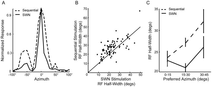
Figure 2.
Receptive field sharpening during SWN stimulation. A) Example spatial RFs measured using the linear array with sequential stimulation (dashed line) and SWN stimulation (solid line). B) RF half-widths decreased for more than 80% of the neurons (above unity line) from the sequential to the SWN stimulation conditions (Wilcoxon signed-rank test, p<0.001). C) RF half-width’s dependence on preferred azimuth for sequential and SWN stimulation. Using sequential stimulation (dashed line), the tuning width increased linearly with preferred azimuth. However, tuning width was not significantly broader among groups in the SWN condition (solid line). RF sizes obtained with sequential stimulation were significantly broader for the intermediate and peripheral groups compared to RFs measured through SWN stimulation (One-way ANOVA, p<0.05). Error bars represent SEM.

