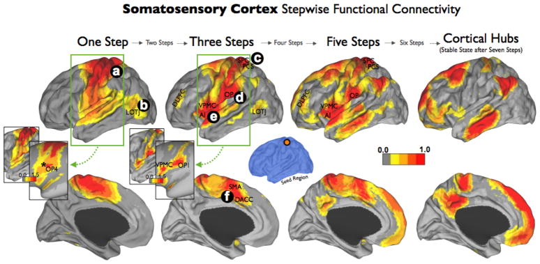
Figure 4. Somatosensory Cortex Stepwise Functional Connectivity.
Somatosensory cortex SFC analysis showed dense connections within the entire somatomotor cortex and a strong direct connectivity with the secondary somatosensory area SII in OP4 (a and star symbol in insert). In order to better visualize the degree of connectivity, conventional (lower image) and inflated projections (upper image) of the results using a relaxed color-scale threshold and centered on the insula region/postcentral gyrus (green square) are presented in the one step and three steps maps. Primary somatosensory cortex had later connectivity to the multimodal network (c, d, e, f) and to the cortical hubs. Labels in cortical maps: LOTJ= Lateral Occipitotemporal Junction; VPMC= Ventral Premotor Cortex; AI= Anterior Insula; OP= Operculum Parietale; OP1= Operculum Parietale 1; OP4= Operculum Parietale 4; SPC= Superior Parietal Cortex; PCS= Posterior Central Sulcus; DLPFC= Dorsolateral Prefrontal Cortex; SMA= Supplementary Motor Area; DACC= Dorsal Anterior Cingulate Cortex.

