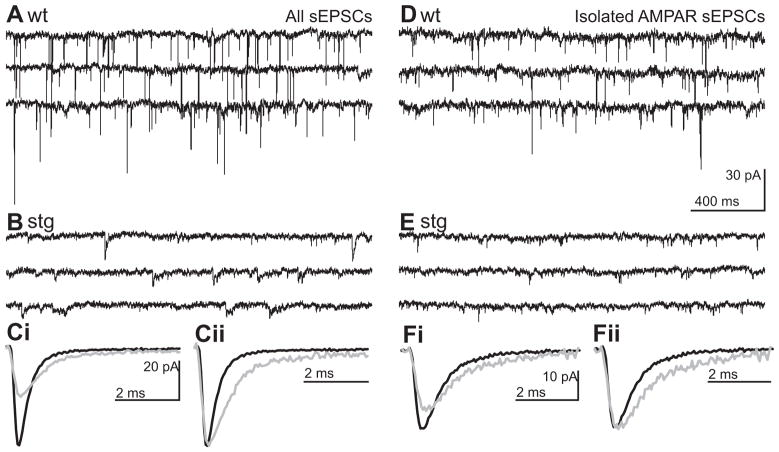
Figure 2. Frequency, amplitude and kinetics of spontaneous EPSCs are altered in stg RTN cells.
Representative traces from wt (A and D) and stg (B and E) RTN cells of continuous voltage-clamp recordings of RTN cells held at resting membrane potential (Vh = −60 mV) under conditions that isolate ionotropic sEPSCs (A–C) and AMPAR sEPSCs (D–F).. Ci Ensemble averaged sEPSCs from representative cells shown in A (black; wt) and B (grey; stg) superimposed on the same time-scale. Cii Averaged sEPSC from Ci normalized and aligned to peak to illustrate the slower kinetics of stg sEPSCs. Di Ensemble averaged sEPSCs from representative cells shown in D (black; wt) and E (grey; stg) superimposed on the same time-scale. Dii Averaged sEPSC from Di normalized and aligned to peak to illustrate the slower kinetics of stg AMPAR-sEPSCs. Remaining events were blocked by NBQX or DNQX 20 μM confirming that the latter were AMPA receptor mediated events (data not shown).

