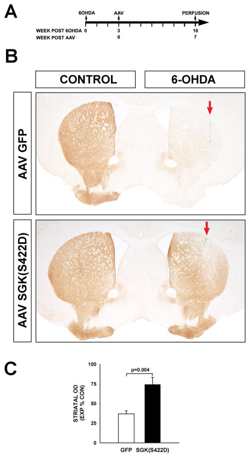
Figure 5. Constitutively active SGK1 induces restoration of the nigro-striatal dopaminergic projection following its destruction by neurotoxin lesion.
A, Mice received an intra-striatal 6-OHDA lesion at Time = 0, and then at 3 weeks post-lesion received AAV hSGK1(S422D) or AAV GFP injection control. B, At 7 weeks following AAV injection, extensive reinnervation of the striatum is observed, as shown in representative coronal sections stained for TH. In each panel, the red arrow indicates the site of the 6-OHDA injection (CONTROL: Contralateral, uninjected side). C, Restoration of striatal innervation is shown quantitatively as optical density of TH staining, with the Experimental (EXP) lesioned side expressed as a percent of density in the Control (CON) non-lesioned side (p=0.004, t-test; AAV GFP N=6, AAV hSGK1(S422D) N=7).

