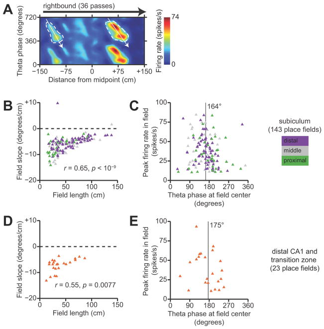
Figure 8.
Theta phase precession in unitary place fields. A, Illustration of the place field segmentation method. A position-phase firing-rate map is shown for a representative neuron in the subiculum. Segmented place fields are outlined in a dashed white contour, and white arrows indicate the principal axis of phase precession within each field. B, Summary of field length and phase precession slope for all unitary place fields of subicular neurons with significant position-phase correlations. Each symbol corresponds to a unitary place field. Symbols are color-coded according to the anatomical location of the neuron along the transverse axis of the subiculum. Phase precessions slope is positively correlated with field length, so that larger place fields have shallower slopes. C, Summary of preferred theta phase and peak firing rate for the same unitary place fields as in B. The vertical line indicates the mean phase for all segmented place fields, which is near to the trough of the LFP theta oscillation. D, Summary of field length and phase precession slope for all unitary place fields of CA1 neurons with significant position-phase correlations. E, Summary of preferred theta phase and peak firing rate for the same unitary place fields as in D.

