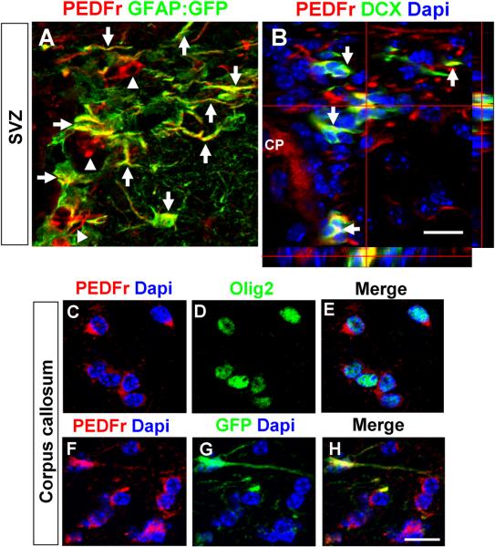
Figure 4. Immunohistochemical analysis of PEDF receptor expression in the adult SVZ and corpus callosum.
(A) Confocal image showing expression of PEDF receptor in GFAP:GFP+ (arrows) and GFAP:GFP− cells (arrowheads) in the SVZ. (B) DCX+ neuroblasts (indicated by arrows and orthogonal view) in the SVZ expressed PEDF receptor. CP: Choroid plexus. Scale b a r = 2 5μm. (C–H) PEDF receptor expression was detected in both Olig2+ oligodendroglial lineage cells (C–E) and GFAP:GFP+ astrocytes (F–H) in the adult corpus callosum. Scale bar = 20μm.

