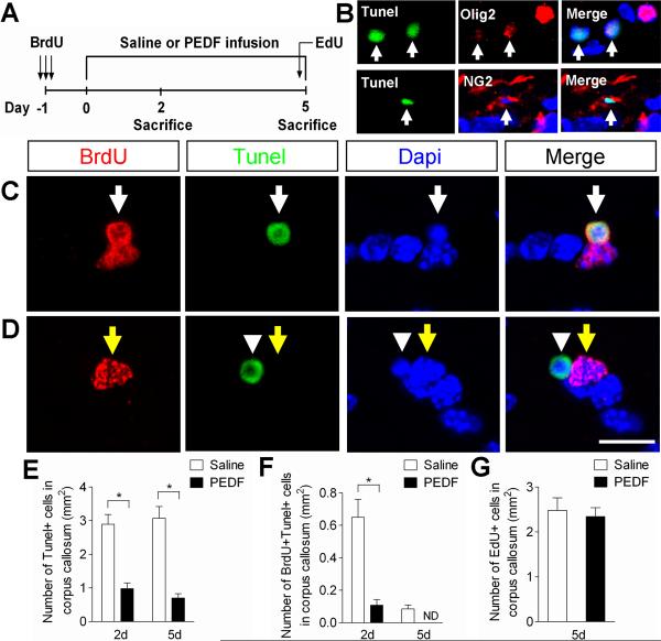
Figure 8. PEDF enhances cell survival in the adult corpus callosum.
(A) Experimental design: adult wild type mice received three consecutive administrations of BrdU (100mg/kg body weight ip) at 6 hour intervals to label cycling OPCs in the corpus callosum, followed by saline- or PEDF- (300ng per day) intracerebral infusion for 2 or 5 days. At day 5, EdU (100mg/body weight ip) was also injected into the animals 5 hours prior to sacrifice. (B) Confocal images of cells co-labeled for Tunel and Olig2 or NG2 in saline-infused corpus callosum, showing oligodendroglial lineage cells undergoing apoptosis. (C–D) Confocal images of cells double-assayed for BrdU and Tunel in the saline- (C) or PEDF- (D) infused corpus callosum. (C) BrdU+/Tunel+ cell is indicated by white arrows. (D) BrdU+/Tunel− cell is indicated by yellow arrows. BrdU−/Tunel+ cell (arrowheads) confirms the absence of cross reactivity between BrdU and Tunel staining. (E–F) Percentages of Tunel+ (E) and BrdU+/Tunel+ (F) cells in saline- or PEDF-infused corpus callosum at day 2 and 5, indicating a potent cell survival effect of PEDF on the corpus callosal cells including newly born OPCs (F). (G) EdU incorporation assay indicating that PEDF did not significantly alter cell proliferation in the corpus callosum. ND = not-detected. Scale bar = 15μm. Results are means +/− SEM (n= 3 brains) (* P<0.01, Student's t test).

