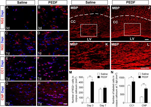
Figure 9. PEDF promotes oligodendroglial regeneration in lysolecithin-induced demyelinative corpus callosum.
Administration of lysolecithin to adult corpus callosum to induce focal demyelination was followed by saline- or PEDF (300ng per day)-infusion for 3 or 7 days. (A–H) Confocal images of cells in saline- or PEDF-infused corpus callosum immunolabeled with antibodies against NG2 (A–D), CC1 (E–F), or CNP (G–H) at 3 or 7 days post-lesion (dpl). Scale bars = 25μm. (I–L) MBP immunostaining of saline- (I and K) or PEDF- (J and L) infused corpus callosum at 7 dpl. (K and L) High magnification images of the areas indicated by rectangles in I and J, respectively. Scale bars = 50μm. (M–N) Quantification for NG2+ OPCs at 3 and 7 dpl (M), and CC1+ and CNP+ mature oligodendrocytes (N) at 7 dpl. Note that PEDF infusion resulted in markedly higher numbers of NG2+ cells present in the lesional corpus callosum at 3 dpl, and substantially enhanced CNP+ mature oligodendrocyte production and CNP immunoreactivity, and MBP positive myelin levels at 7 dpl. CC: corpus callosum. LV: lumen of lateral ventricles. Results are means +/− SEM (n = 4 brains, * P<0.05, and ** P<0.01, Student's t test).

