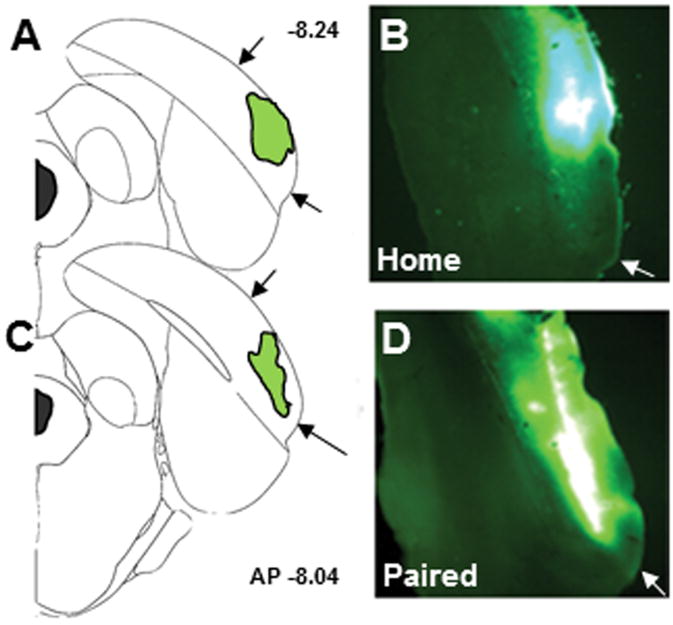
Figure 4.
Representative images of the placement of infusions of the retrograde tracer CTb-488 (cholera toxin subunit B conjugated to AlexaFluor 488) into POR. (A, C) Brain diagrams depicting infusion locations within POR. The anteroposterior (AP) location is noted to the right of the brain diagrams. (B, D) Images of the fluorescent label indicating correct POR placement. Black arrows in A and C mark the dorsoventral boundaries of POR and white arrows in B and D mark the ventral boundary of POR.

