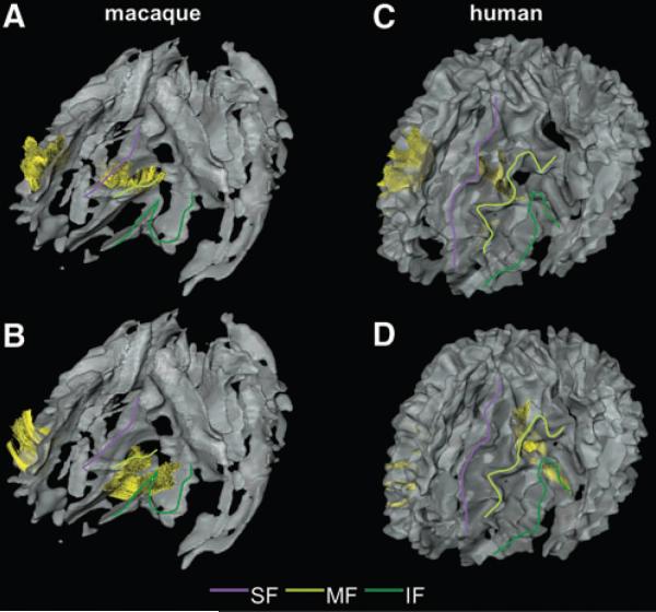
FIG. 3.
The U-fibers in the frontal area of macaque (A and B) and human (C and D) brains. U-fibers are displayed as yellow fibers. Color lines are drawn on each blade of the left side of the brain to help the visual identification: purple line, SF; light green line, MF; and deep green line, IF. (A and C) The U-fibers connecting the SF and the MFs. (B and D) The U-fibers connecting the MF and the IFs.

