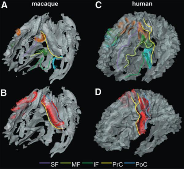
FIG. 4.
The U-fibers in the central area of the macaque (A and B) and human (C and D) brains. Color lines are drawn on each blade of the left side of the brain to help the visual identification: purple line, SF; light green line, MF; deep green line, IF; yellow line, PrC; and blue line, PoC. (A and C) The U-fibers connecting the SF and the PrCs (orange fibers); the U-fibers connecting the MF and the PrCs (green fibers); and the U-fibers connecting the IF and the PrC (light blue fibers) are shown. (B and D) The U-fibers connecting the PrC and the PoC (red fibers) are shown.

