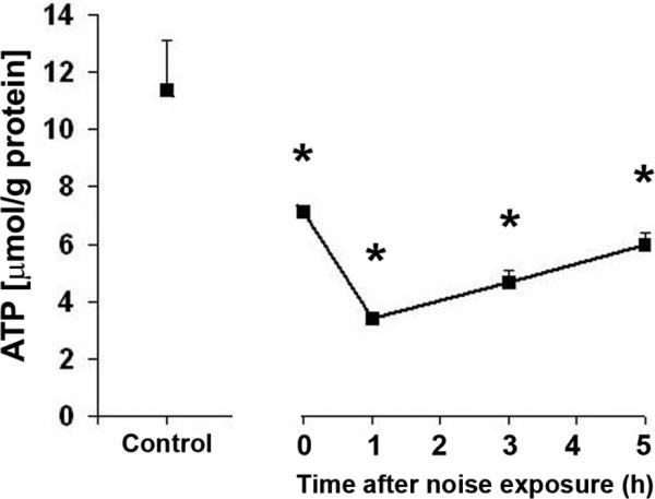
Figure 3.

ATP levels in the cochlear tissue of the inner ear: The level of ATP decreased significantly immediately after noise exposure and was lowest at 1 h post-noise exposure. The ATP level recovered slowly, but remained significantly lower than those of control mice at 5 h post-noise exposure. Data are presented as means + SD; *p < 0.05, n = 7.
