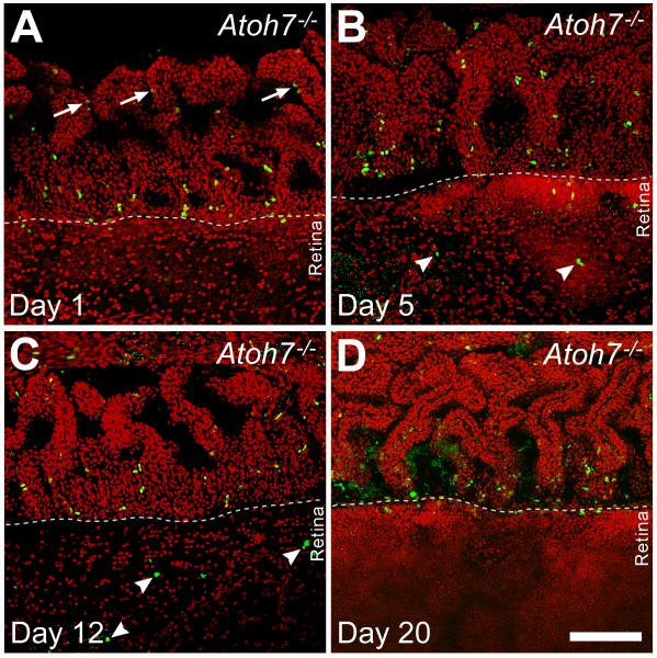
Figure 5.
Confocal images of flat mounted retinas from Atoh7-deficient (95% RGC depleted) mice pulse labeled with BrdU at P15. In all panels, the ciliary margin is in the upper half and retinal margin is in the lower half. A. One day after injection, BrdU positive cells are most abundant in a line at the planoretinal junction. Additional labeled cells are present in the pars plana closer to the cilia, where many appear in pairs (arrows) with weaker BrdU signals suggesting continued cell division. B. Five days after injection, fewer BrdU-positive cells remain at the planoretinal junction but strongly-labeled BrdU positive cells are present in both the pars plana and retina suggesting cells have migrated away from the planoretinal junction. Small, punctate BrdU residues are also visible in the retina suggesting possible cell death among migrating cells. C. Twelve days after injection, fewer BrdU positive cells can be observed in either the pars plana or retina. BrdU positive cells (arrowheads) can be observed further inside the retina. However, many of these cell nuclei show irregular shapes and there are additional sites of punctate staining suggesting possible apoptosis. D. Twenty days after injection, increasing photon multiplier gain is needed to detect cells with weak BrdU staining at the planoretinal junction, suggesting that the few remaining cells have gone through additional rounds of mitosis to dilute the BrdU following the initial labeling. Nuclear debris indicated by diffuse, punctate BrdU staining can also be seen at the junction. Scale bar = 100 μm.

