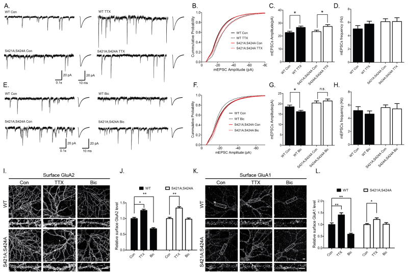
Figure 1. Mecp2S421A;S424A/y neurons exhibit intact TTX-induced synaptic scaling up, but impaired bicuculline-induced synaptic scaling down.
A) Representative whole-cell recording sample traces, average mEPSC waveforms from cultured Mecp2S421A;S424A/y and wild type hippocampal neurons (18 DIV) treated with either control solution or TTX (1 μM) for 48h–72h. B) Cumulative probability distribution of mEPSC amplitude from cultured Mecp2S421A;S424A/y and wild type hippocampal neurons (18 DIV) treated with either control solution or TTX (1 μM) for 48h–72h. C) Quantification of mean mEPSC amplitude for each population in A (n=49–54 cells each group). D) Quantification of mean mEPSC frequencies for each population in A. E) Representative whole-cell recording sample traces, average mEPSC waveforms from cultured Mecp2S421A;S424A/y and wild type hippocampal neurons (18 DIV) treated with control solution or bicuculline (40 μM) for 48h–72h. F) Cumulative probability distribution of mEPSC amplitude from cultured Mecp2S421A;S424A/y and wild type hippocampal neurons (18 DIV) treated with control solution or bicuculline (40 μM) for 48h–72h. G) Quantification of mean mEPSC amplitude for each population in E (n=47–61cells each group). H) Quantification of mean mEPSC frequencies for each population in E. I–L) Representative images and quantification of surface GluA2 and GluA1 immunoreactivity in Mecp2S421A;S424A/y and wild type cultured hippocampal neurons (18 DIV) treated with control solution, TTX (1 μM) or bicuculline (40 μM) for 48h. Scale bar: 10 μm. (n=15–20 cells each group). The bar graph shows the mean ± s.e.m * p<0.05. ** p<0.01

