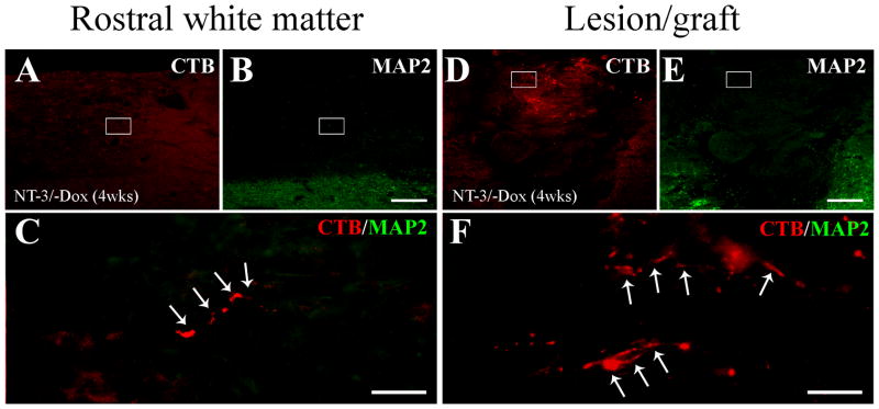
Figure 12. Regenerated sensory axons do not form axodendritic contacts.
Double immunolabeling of CTB-labeled regenerated sensory axons (red, arrows) and MAP2-labeled dendrites (green) (A–C) rostral to the lesion site and (D–F) within the lesion site indicates a lack of dendrites in the vicinity of regenerated axons after 4 weeks of NT-3 expression. (C) and (F) are high magnification of boxed regions in (A, B) and (D, E), respectively. Scale bars: 200 μm in (A, B, D, E); 50 μm in (C, F).

