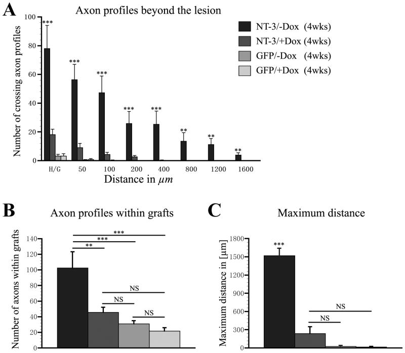
Figure 7. Quantification of regenerated CTB-labeled sensory axon profiles within and rostral to the lesion site.
(A) The number of axonal profiles crossing a virtual line at the rostral host/graft interface (H/G, 0 μm) and at different distances beyond the interface (50–1600 μm) was quantified in one out of seven sagittal sections. Animals that received tet-off-NT-3 virus and received no doxycycline (−Dox) exhibit significantly more axonal profiles beyond the rostral lesion border than animals that were treated with doxycycline to turn NT-3 expression off (+Dox) and control animals that received GFP virus with or without doxycycline treatment. (B) Similarly, significantly more axonal profiles are present within cellular grafts in animals that received tet-off NT-3 virus when gene expression is turned on compared to all other groups. (C) The maximum average distance of axon growth is also significantly higher in animals that received tet-off NT-3 virus when gene expression is turned on (**p < 0.01, ***p < 0.001, NS p > 0.05; ANOVA followed by Fisher’s post hoc test).

