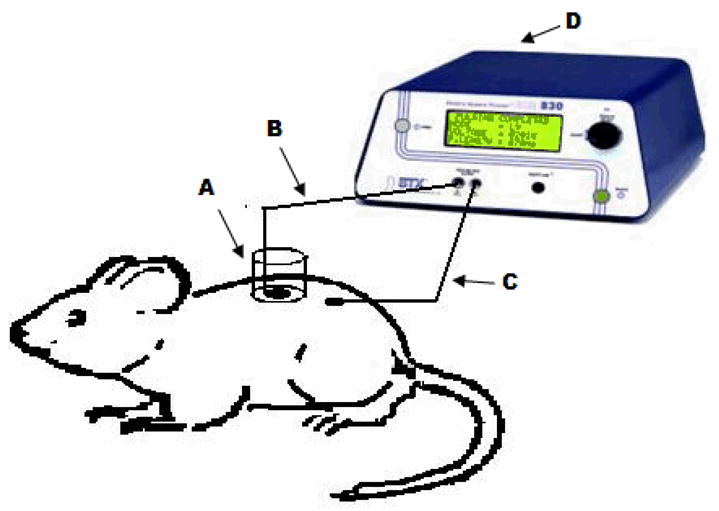
Figure 1.

Diagram representing the in vivo experimental set up is shown in this figure. The sampling chamber (A) was glued on the skin surface of the sprague dawley rat. Ag-Agcl electrodes (B and C) are placed in the sampling chamber and secured on the skin surface respectively. 0.4 ml of sampling buffer was placed in the chamber. The two electrodes were connected to BTX 830M electrosquare porator and electrical pulses were applied. The sampling buffer was collected after 15 minute and the amount of glucose present was measured.
