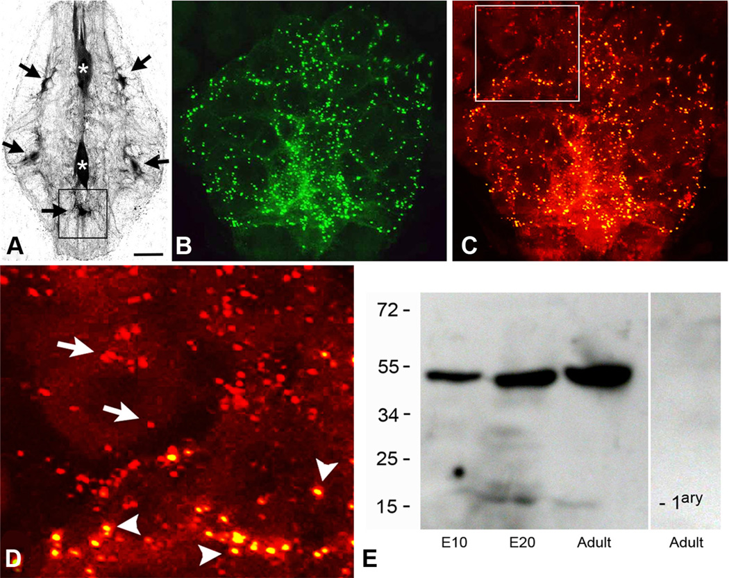
Figure 3.
The punctal distribution of the INX2-EGFP expression in ganglionic glial cells closely resembles the punctal distribution of the endogenous INX2 protein.A, The large glial cells in the leech ganglion are electrically coupled and tracer coupled. Neurobiotin injection into any one of them leads to labeling of all the cells. Fluorescent streptavidin labeling of a leech ganglion is shown here after the anterior neuropile glial cell (top asterisk) was injected with Neurobiotin. Strong streptavidin labeling can be seen in the posterior neuropile glial cell (bottom asterisk), as well as, five of the packet glial (PG) cells (black arrows). B, INX2-EGFP transgene expression in the posterior ventral medial PG cell (corresponding approximately with the location indicated by the box in A). A punctate pattern of staining can be seen distributed throughout the processes of the cell, which surround and outline many of the neuronal cell bodies. C, INX2 antibody staining (red) of the same cell and region as shown in B. D, Close-up view of the boxed area in C, showing the double labeling of the INX2 transgene (yellow spots, arrowheads) and the single labeling of the endogenous gap junctions (red, arrows) in adjacent glia, outside the boundary of the expressing cell. E, INX2 SDS-PAGE immunoblot analysis of leech lysates. From the left, the first lane shows lysate from an early embryo (E10); the second, a late embryo (E20); and the third, the adult CNS. The antiserum in each recognizes a band a little below 55 kDa. The lane on the right-hand side is the adult lysate with secondary antibody only. Scale bars: A, 75µm; B, C, 15µm; D, 7µm.

