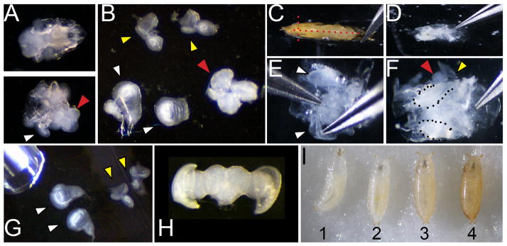
Figure 1. Dissection and staging of larval and pupal tissues.

A. Dissection of larvae (of genotype w1118 shown) at 110 hours of development showing the anterior third before (top) and after (bottom) inversion. White arrowhead indicates a wing attached to the larval cuticle and a red arrowhead indicates the position of the brain B. Wings (white arrowhead; note wing at left is attached to a leg disc, wing at right is torn at the dorsal notum), eyes (yellow) and brain (red) removed from the larvae. C. Dissection of pupa showing positions of cuts with forceps placed at the airspace anterior to the head (red dotted lines) D. Pupa removed from cuticle before washing to remove fat and gut. E. Cleaned pupa showing process of lifting wings for removal F. Cleaned pupa showing wings (dotted) and partially removed eye-brain complex (red and yellow arrowheads). G. Dissected larval wings and eyes showing pipette for transfer. H. Dissected post-mitotic pupal eye-brain complex. I. Correct staging of animals at the larval/prepupa transition; animal 1 is still moving and is too young; animal 2 is at the correct stage to collect, 0hAPF (after pupa formation) it is white and immobile; animal 3 is too old to collect, the darkening of cuticle is evident by 1hAPF; animal 4 is too old to collect already tanned at 2hAPF.
