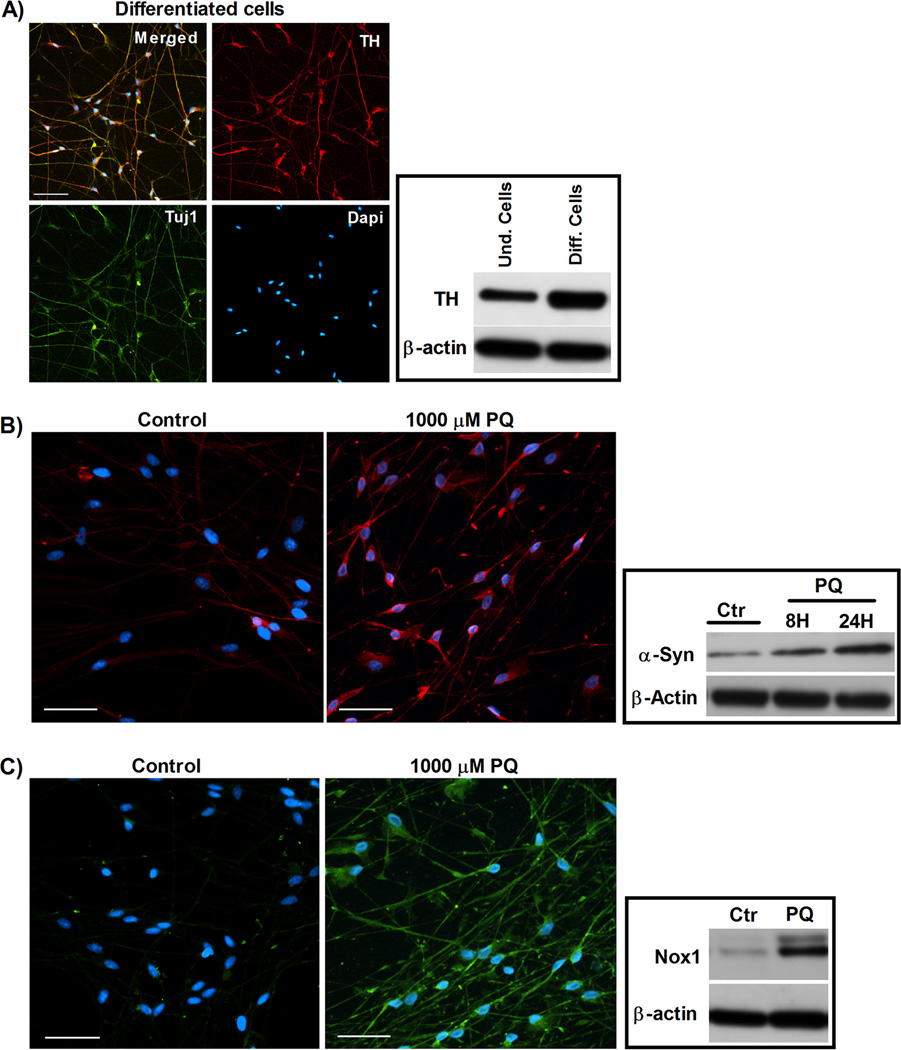
FIG. 1. Increases in α-synuclein and Nox1 in human dopaminergic neurons exposed to PQ.
(A) Characterization of human ventral mesencephalic neuronal progenitor cell line, ReNcell VM, after differentiation (human dopaminergic neurons). Left panel depict representative photomicrographs of TH, Tuj1 and DAPI immunostaining of ReNcell VM after 14-day differentiation. The right panel displays the expression of TH protein in ReNcell VM, before and after differentiation. (B) α-Synuclein levels in differentiated human dopaminergic cells exposed to PQ. Left panel shows α-synuclein immunoreactivity (red). Right panel represent α-synuclein protein levels in immunoblot. (C) Nox1 levels in differentiated human dopaminergic cells exposed 8H to PQ. Left panel shows Nox1 immunoreactivity (green). Right panel illustrates Nox1 protein levels in immunoblot. β-actin was used as an internal control. Und: undifferentiated and Diff: differentiated. Ctr: control and PQ: paraquat. Scale bars = 50 µm.

