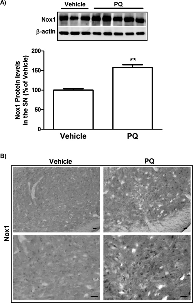
FIG. 6. Increase in Nox1 protein levels in the SN of rats injected with PQ.
(A) Representative immunoblot and quantitative analysis of Nox1 protein levels. Nox1 protein was determined in the total lysates of SN tissues of rats injected with vehicle or PQ by immunoblot analysis. β-actin was used as an internal control. PQ significantly increased Nox1 protein that was quantified using Quantity One software and normalized against β-actin. (B) Representative photomicrographs of Nox1-immunoreactivity in the SN sections of rats injected with vehicle or PQ. Nox1 immunoreactivity in the SN was increased in PQ injected animals compared to vehicle. The result is expressed as percentage of vehicle. Data are shown as the mean ± SEM. Statistical analysis was performed using the Student t test. **P<0.01. Scale bars = 50 µm.

