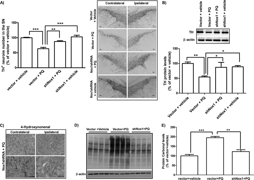
FIG. 8. Nox1 knockdown reduced SN dopaminergic neuronal death induced in rats administered with PQ.
(A) Representative photomicrographs of TH-immunostaining and quantitative analysis of TH-positive dopaminergic neurons in the SN of rats after Nox1 knockdown. Representative photomicrographs of TH-immunoreactivity in the SN of the contralateral and ipsilateral sides of brain sections of the four experimental groups. TH-positive neurons in the ipsilateral side were stereologically counted. (B) Representative immunoblot and quantitative analysis of TH protein levels. TH protein was determined in total lysates of the rats SN tissues in the ipsilateral side by immunoblot analysis β-actin was used as an internal control. TH protein levels were quantified using Quantity One software and normalized against β-actin. (C) Representative photomicrographs of 4HNE immunostaining in the SN of the contralateral and ipsilateral sides of brain sections of rats from shNox1 + PQ group. Scale bars = 50 µm. (D and E) Immunoblot (D) and quantitative analysis (F) of protein carbonyl levels determined in total lysates of rats ipsilateral SN tissues. β-actin was used as an internal control. The results are expressed as percentage of vector + vehicle. Data are shown as the mean ± SEM. Statistical analysis was performed using one-way ANOVA followed by Bonferroni’s Multiple Comparison Test. *P<0.05; **P<0.01 and ***P<0.001.

