Figure 2.

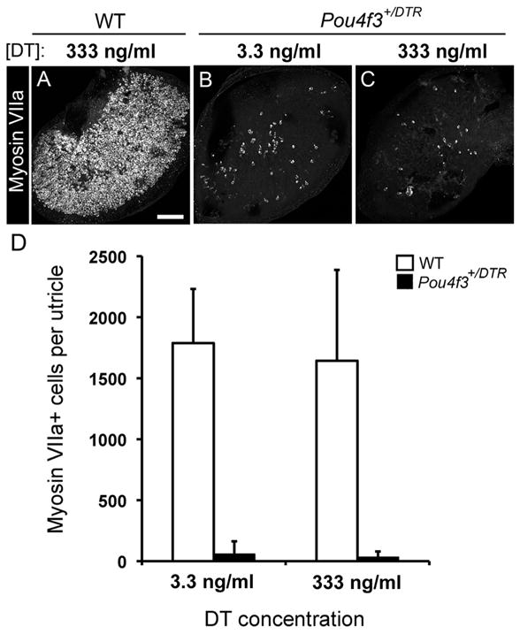
Hair cell loss is increased but not complete after in vitro DT treatment. Hair cell loss was assessed qualitatively and quantitatively in adult utricles that were cultured with DT for 24 h and for 5 additional days in DT-free media. A–C, Confocal brightest point projection images of whole-mount utricles labeled for myosin VIIa from wild-type (WT) mice treated with 333 ng/ml DT (A) or from Pou4f3+/DTR mice treated with either 3.3 ng/ml (B) or 333 ng/ml (C) DT. Scale bar: (in A) A–C, 100 μm. D, Graph shows mean numbers of myosin VIIa+ cells (±SD) per utricle for wild-type mice treated with 3.3 ng/ml DT (n = 3 utricles) or 333 ng/ml DT (n = 3 utricles) and for Pou4f3+/DTR mice treated with 3.3 ng/ml DT (n = 5 utricles) or 333 ng/ml DT (n = 8 utricles).
