Figure 4.

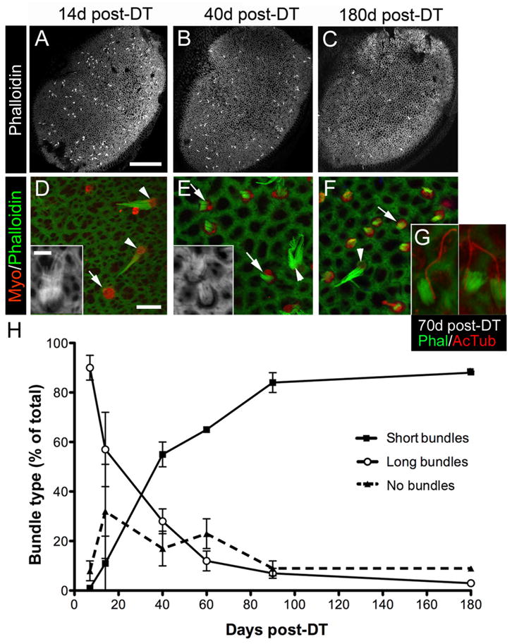
Bundle morphologies on replacement hair cells following DT treatment. A–C, Confocal images of phalloidin labeling at the lumenal surface of utricles at 14 days (d) (A), 40 days (B), and 180 days (C) post-DT. Long bundles of presumed original hair cells, appearing as white dots or dashes, are distributed throughout the sensory epithelium. D–F, Confocal slices near the lumenal surface in the presumed lateral extrastriola from utricles labeled for myosin VIIa (Myo) and phalloidin to detect hair cells and bundles, respectively. At 14 days post-DT (D), myosin VIIa+ hair cells with no bundle (arrow) or a long bundle (arrowheads and inset) were predominant. At 40 days (E) and 180 days (F) post-DT, myosin VIIa+ hair cells with a short bundle (arrows and inset) were most common, but hair cells with long bundles (arrowheads) were also present. G, Three examples of replacement hair cells with a distinct, eccentrically located kinocilium [labeled by anti-acetylated tubulin (AcTub) antibodies] on the side of each bundle [labeled by phalloidin (Phal)] at 70 days post-DT. H, Graph shows mean bundle type, expressed as the percentage (%) of total myosin VIIa+ hair cells (±SD), in utricles from Pou4f3+/DTR mice at several times post-DT. Sample numbers for counts are provided in Table 1. Scale bar: (in A) A–C, 100 μm; (in D) D–F, 6 μm; (in D inset) D inset, E inset, and G, 3 μm.
