
Figure 5.
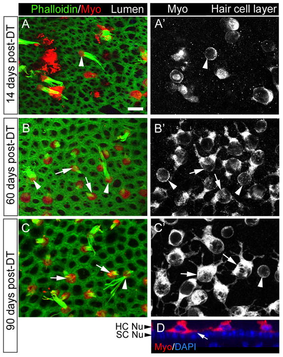
Shapes of replacement hair cells following DT treatment. A–C′, Three sets of confocal slices taken near the lumenal surface in the presumed lateral extrastriola (A, B, C, labeled for phalloidin and myosin VIIa or Myo) or at the level of the hair cell bodies (A′, B′, C′, labeled for Myo). A, A′, The same field at 14 days post-DT. B, B′, The same field at 60 days post-DT. C, C′, The same field at 90 days post-DT. Arrowheads point to cells with long bundles and round cell bodies (presumed original hair cells), while arrows point to cells with short bundles and multipolar cell bodies (presumed replacement hair cells). D. Vertical slice through the presumed lateral extrastriola at 90 days post-DT, illustrating the location of the hair cell processes above the supporting cell nuclear layer (SC Nu). HC Nu, Hair cell nuclear layer. Scale bar (in A) A–D, 6 μm.