Figure 9.

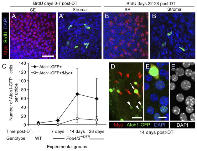
In the absence of supporting cell division, Atoh1 is transcriptionally activated in DT-treated utricles. A–B′, BrdU labeling in damaged utricles. Virtually no BrdU uptake was seen in the sensory epithelium (SE) in Pou4f3 +/DTR utricles after DT treatment. Confocal images show the utricular sensory epithelium (SE) or stroma from Pou4f3 +/DTR mice that received BrdU between 0 and 7 days post-DT (A, A′) or between 22 and 28 days post-DT (B, B′). Myosin VIIa (Myo) labeling is shown in red, BrdU labeling is in green, and DAPI (nuclear) labeling is in blue. C–E′, Atoh1 transcriptional activity in the damaged utricle. C, Graph of the mean number of Atoh1-GFP+ and Atoh1-GFP+/myosin VIIa + cells per utricle (±SD) for the following experimental groups: uninjected (−) wild-types (WT) (n = 5 utricles) and Pou4f3 +/DTR mice at 7 days post-DT (4 utricles), 14 days post-DT (4 utricles), and 28 days post-DT (5 utricles). D, Atoh1-GFP+ (green) nuclei at 14 days post-DT in cells that were either myosin VIIa+ (arrow, red cytoplasm) or myosin VIIa− (arrowheads). E, E′, A higher magnification of the Atoh1-GFP+/myosin VIIa+ cell indicated by the arrow in D. E, Labeling for Atoh1-GFP (green), myosin VIIa (red), and DAPI (blue). E′, DAPI used only to illustrate nuclear localization of the Atoh1-GFP in E. Scale bar (in A) A–B′, 24 μm; (in D), 10 μm; (in E) E, E′, 4 μm.
