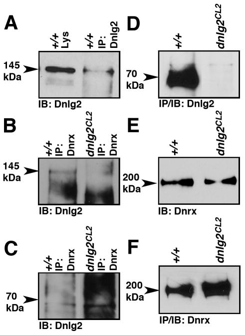
Figure 5. Dnlg2 forms a biochemical complex with Dnrx.
(A) IPs from wild type fly head lysates using anti-Dnlg2 antibodies show the presence of Dnlg2 (145 kDa, arrowhead).
(B, C) IP from wild type fly head lysates using anti-Dnrx reveals the presence of Dnlg2 in the same complex (B, 145kDa, arrowhead). The 70kDa Dnlg2 does not associate with Dnrx. Only non-specific background bands are observed in the wild type and dnlg2 lysates near where the 70kDa band is expected (C, arrowhead). Note that panels B and C are from the same protein blot probed separately.
(D) IPs from equal amounts of wild type and dnlg2 mutant fly head lysates using anti-Dnlg2 show presence of Dnlg2 (70kDa) in wild type but not in dnlg2 mutants. (Note the break in between the lanes is due to removal of an empty lane).
(E) Dnrx protein levels are unaffected in dnlg2 mutants.
(F) IPs from equal amounts of wild type and dnlg2 mutants using anti-Dnrx show the presence of Dnrx in both wild type and dnlg2 mutants.

