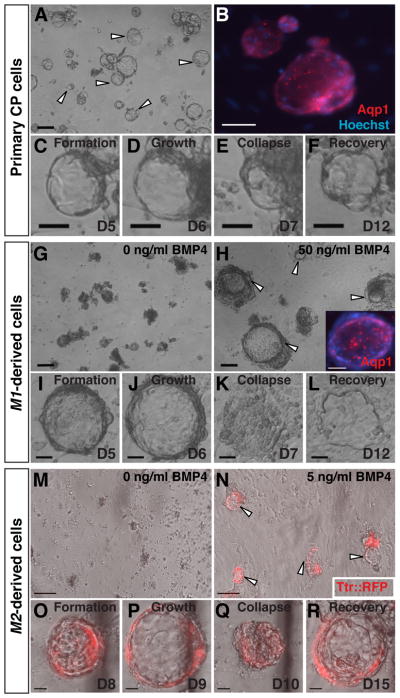
Fig. 4. Vesicle self-assembly and secretion by primary and derived CPECs.
(ICC, phase contrast, and fluorescence microscopy; the same vesicle is shown in C–F, I–L, or O–R).
(A–F) Primary CPECs. CPECs from dissociated primary CP self-assemble into vesicles on Matrigel (arrowheads in A) that are Aqp1-positive (B). Due to their secretory activity, these vesicles enlarge (C,D), collapse upon treatment with the secretion inhibitors acetazolamide and ouabain (E), then regrow upon inhibitor withdrawal (F). (G–L) M1-derived cells. M1 aggregates treated with 50 ng/ml BMP4 (n=6/6 cultures), but not controls (n=0/6 cultures), formed Aqp1-positive vesicles after dissociation and plating on Matrigel (G,H). Like primary CPEC vesicles, M1-derived vesicles expand (I,J), collapse upon secretion inhibitor treatment (K), and recover after inhibitor withdrawal (L). (M–R) M2-derived cells. M2 aggregates treated with 5 ng/ml BMP4 (n=3/3 cultures), but not controls (n=0/3 cultures), form TTR::RFP-expressing vesicles (N) that expand, collapse upon secretion inhibitor treatment, and regrow after inhibitor withdrawal (O–R) in a fashion indistinguishable from primary CPECs. Scale bars: A–N, 100 um; O–R, 50 um.

