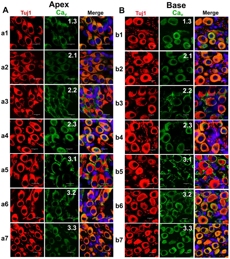
Figure 12. Expression of Cav1.3, Cav2.1, Cav2.2, Cav2.3, Cav3.1, Cav3.2, and Cav3.3 in mouse cochlea sections.
A–B. To rule out the possibility that both apical (A) and basal (B) SGNs expressed Ca2+ channels in vivo when compared to the cell culture-induced expression of channels, we performed immunolabeling as described in Figure 10 on sections of the cochlea from a 3-month-old mice. Apical spiral ganglion neurons were labeled with antibodies against Cav1.3, Cav2.1, Cav2.2, Cav2.3, Cav3.1, Cav3.2, and Cav3.3. As shown, neurons were labeled with the neuronal marker Tuj1 (in red), the channels (in green), and the nuclei were stained with DAPI (in blue) and the merged images (right panel). Spiral ganglion neurons stained positively to Cav1.3, Cav2.1, Cav2.2, Cav2.3, Cav3.1, Cav3.2, and Cav3.3. B Similar data were obtained for basal neurons as shown for Cav1.3, Cav2.1, Cav2.2, Cav2.3, Cav3.1, Cav3.2, and Cav3.3, respectively. Scale bar = 10 μm.

