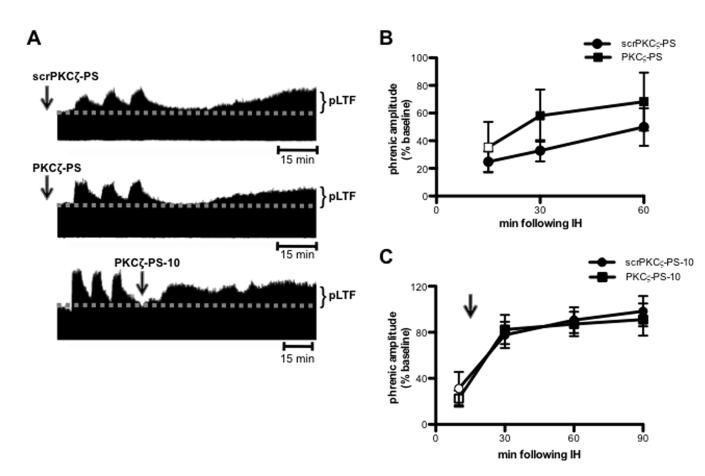
Figure 3.
Intermittent hypoxia induced pLTF does not require spinal aPKC activity. A. Representative compressed and integrated phrenic neurograms before, during and for 60-90 min following 3, 5 min episodes of hypoxia (11% O2), illustrating the development of pLTF in rats receiving intrathecal scrPKCζ-PS (top) or PKCζ-PS (middle) 20 min prior to IH, or PKCζ-PS delivered 10 min (PKCζ-PS-10; bottom) following IH. Arrows indicate time points of drug delivery. B. Average change in phrenic burst amplitude from baseline for 60 min following IH in rats receiving intrathecal scrPKCζ-PS (circles) or PKCζ-PS (squares) 20 min prior to IH. Both rat groups exhibited significantly increased phrenic burst amplitude 60 min following IH relative to baseline. No differences were observed between rats receiving intrathecal scrPKCζ-PS or PKCζ-PS at any point. C. Average change in phrenic burst amplitude from baseline following IH in rats receiving intrathecal scrPKCζ-PS (circles) or PKCζ-PS (squares) 10 min following IH. Black arrow indicates time point of drug delivery. Both groups exhibited significant increases in phrenic burst amplitude at 30, 60 and 90 min following IH relative to baseline. No differences were observed between rats receiving intrathecal scrPKCζ-PS-10 or PKCζ-PS-10. Mean values + SEM. Filled symbols indicate significantly different than baseline. p<0.05

