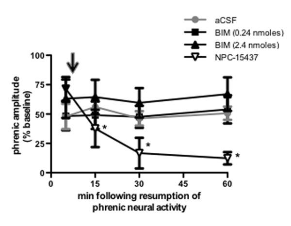
Figure 4.
Stabilization of iPMF requires activity of spinal PKCζ/Ι, but not classical or novel PKC isoforms. Average change in phrenic burst amplitude from baseline for 60 min following neural apnea in rats receiving intrathecal NPC-15437 (inverted triangles; inhibits PKC isoforms with a regulatory subunit), 0.24 nmoles BIM (squares; novel and classical PKC inhibitor) or 2.4 nmoles BIM (triangle) 10 min after resumption of respiratory neural activity. Black arrow indicates time point of drug delivery. All rats expressed significant increases in phrenic burst amplitude relative to baseline or time controls (not shown) immediately prior to drug injections, indicating iPMF. iPMF progressively declined following NPC-15437 injections; by 15 min following resumption of respiratory neural activity, iPMF was significantly decreased from pre-injection value and no longer significantly different from baseline or time controls. No change in iPMF magnitude was observed following intrathecal BIM (0.24 or 2.4 nmoles). Rats receiving intrathecal aCSF prior to neural apnea (from figure 1) are shown in gray circles for comparison. Mean values + SEM. Filled symbols indicate significantly different from baseline. *significantly different from pre-injection time point. p<0.05

