Figure 3.

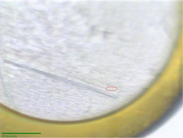
L-selenomethioine crystals mounted on the microdiffractometer MD2 at beam line X06SA. The red ellipsoid corresponds to the focused beam size of 25 × 6 μm (Full width at half maximum).

L-selenomethioine crystals mounted on the microdiffractometer MD2 at beam line X06SA. The red ellipsoid corresponds to the focused beam size of 25 × 6 μm (Full width at half maximum).