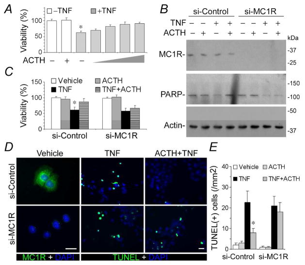
Figure 7. MC1R mediates the anti-apoptotic effect of ACTH in renal tubular epithelial cells.
(A) ACTH reinstates the viability of TNF injured IRPTC cells in a dose dependent fashion. IRPTC cells were treated with vehicle, TNF (10ng/ml), ACTH (10−8M), or TNF in combination with ACTH (10−10M, 10−9M, 10−8M, 10−7M) for 6 h before cellular viability was estimated by MTT assay; *P<0.05 versus other groups. (B) After MC1R silencing, ACTH fails to prevent TNF induced PARP cleavage. IRPTC cells were transfected with scrambled (si-Control) or MC1R specific (si-MC1R) siRNA for RNA interference. Cells were then treated with vehicle, TNF (10ng/ml) and/or ACTH (10−8M) for 6 h. Cell lysates were subjected to immunoblot analysis for MC1R, PARP and actin (B); cellular viability assessed by MTT assay (C); *P<0.05 versus other groups in si-Control. (D) Fixed cells were prepared for TUNEL staining (green), fluorescent immunocytochemistry staining for MC1R (green) and counterstaining of DAPI (blue); scale bar=30μm. (E) Absolute counting of TUNEL positive cells in cell cultures; *P<0.05 versus other groups in si-Control.

