Figure 1.
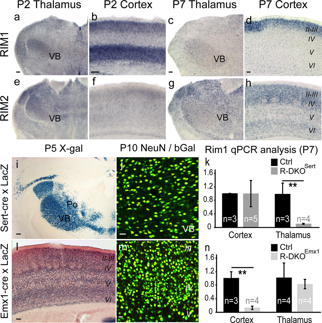
RIM1 and RIM2 expression and Cre-mediated recombination. a–h, In situ hybridization using RIM1 and RIM2 antisense probes at P2 and P7. a–d, RIM1 is expressed in the somatosensory thalamus (VB; a) and in the cortex with highest expression in deep layers at P2 (b) and upper layers at P7 (d). e–h, RIM2 is broadly expressed in the thalamus including the VB (e), with a low expression in the P2 cerebral cortex (f) and higher expression at P7 (h). i–n, Recombination induced by Sert-cre and Emx1-cre. Cre mice were crossed with a reporter strain expressing a nuclear β-Galactosidase (Taumgfp-nls-LacZ). This recombination, visualized by X-gal staining (i, l) and β-Gal/NeuN double immunolabeling (j,m) shows extensive neuronal recombination in the VB of Sert-cre mice (i, j), and cerebral cortex of Emx1-cre mice (l, m). k, n, Thalamic and cortical mRNAs from P7 brains of both strains were extracted and measured by qPCR. Results for Rim1 are summarized in kfor RIM-DKOSert and in nfor RIM-DKOEmx1. **p < 0.05. Scale bars: (in a–i, l, 100 µm; j,m, 25 µm.

