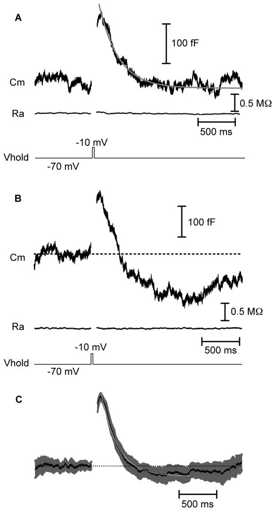
Figure 1. Fast endocytosis in cones.
Whole-cell capacitance recordings from cones in a salamander retinal slice preparation. A) A 25 ms step depolarization to −10 mV from a holding potential of −70 mV evoked an increase in membrane capacitance from fusion of synaptic vesicles. The capacitance trace returned to baseline indicative of endocytosis. The decline of the capacitance trace could be fit with a single exponential function with a time constant (τ) of 257 ms. The baseline access resistance (Ra) was 32.5 MΩ and the baseline membrane capacitance (Cm) was 32.1 pF. The displayed trace is an average of three traces from a single cone. B) As in A, capacitance response to a 25 ms depolarization. In this recording, the capacitance trace overshot the baseline, reflecting excess endocytosis. Baseline values: Cm = 36.3 pF, Ra = 37.9 MΩ. C) Mean (black) and SEM (gray) of 41 normalized capacitance traces fit with a single exponential (white, τ = 221 ms). Cm, membrane capacitance; Ra, access resistance Vhold, holding potential.

